Cell Imaging Highspeed with minimal Photobleaching Biology Diagrams In this long history of cell biology of mitosis, however, whole-cell three-dimensional (3D) live imaging with sufficient spatiotemporal resolution to discriminate nanoscale-sized mitotic machineries and completely track their movements has never been achieved. Liu Z, Lavis LD, Betzig E (2015) Imaging live-cell dynamics and structure at the
73.1K Views. Mitosis is a form of cell division in which a cell's genetic material is divided equally between two daughter cells. Mitosis can be broken down into six phases, during each of which the cell's components, such as its chromosomes, show visually distinct characteristics. Advances in fluorescence live cell imaging have allowed scientists to study this process in great detail Mitosis is the cell cycle phase where the cell divides into two daughter cells. A successful mitosis requires the equal partitioning of the genetic material into two daughter cells. Since then, live-cell imaging has been the method of choice to capture the rapid sequence of mitotic events and the dynamicity of the mitotic spindle. Indeed Live cell imaging was used to visualize the fate of individual cells as they progressed from NEBD through mitosis . Nocodazole is known to arrest the wild type cells at G2M phase by interfering with microtubule formation. It was observed that post nocodazole treatment wild type cells were arrested at metaphase upper panel .
cell imaging at different timescales Biology Diagrams
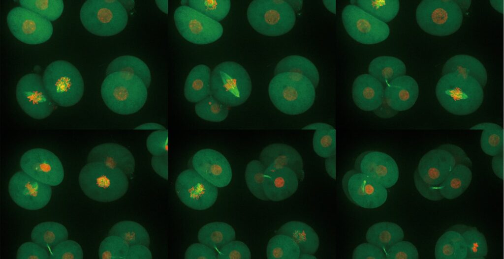
Mitosis is a highly dynamic and choreographed process in which chromosomes are captured by the mitotic spindle and physically segregated into the two daughter cells to ensure faithful transmission of the genetic material. Live-cell fluorescence microscopy enables these dynamics to be analyzed over diverse temporal scales.

1.2 Live-Cell Imaging Considerations 1.2.1 Environment. Maintaining cell health is critical in live-imaging experiments. Mammalian cells must be viable and healthy during the imaging process to ensure that they will progress normally through the cell cycle. Important parameters that must be maintained include temperature, osmotic pressure and pH.
Imaging mitotic processes in three dimensions with lattice ... Biology Diagrams
By comparing mitosis in live wild-type and mutant cells, it has been shown, e.g., that cytoplasmic dynein is required for positioning the spindle in yeast , for the information lost in the undersampled data sets acquired during live cell imaging. This means that such methods can improve the resolution of the original fluorescence image and
Live-cell fluorescence microscopy is a powerful tool for characterizing aberrant mitotic phenotypes resulting from exposure to chemical inhibitors or after depletion of protein targets by RNA interference or other methods. Live imaging of cultured cells during mitotic progression presents challenges …
