Induced Cell Cycle Arrest in TripleNegative Breast Cancer Biology Diagrams In preclinical studies, it was reported to induce cell cycle arrest and tumor growth inhibition in a majority of solid tumor cell lines especially those of B-cell origin and xenografts 88. phase II clinical trials were completed with flavopiridol in chronic lymphocytic leukemia 89, endometrial adenocarcinoma 90, multiple myeloma 91, and
Generating excessive levels of DNA damage, through either radiotherapy or chemotherapy, The cell-cycle arrest and apoptotic functions of p53 in tumor initiation and progression.
Targeting cell cycle and apoptosis to overcome chemotherapy ... Biology Diagrams
We therefore investigated whether therapeutically targeting cell cycle control pathways that converge on the G 1 /S restriction point could synergise with cytotoxic chemotherapy, potentially circumventing chemoresistance. The addition of MDM2 inhibitor, nutlin-3a with AraC/Dox achieved striking synergism in TP53-competent cell lines in reducing viability and promoting apoptosis.
Furthermore, the formation of doxorubicin-DNA adducts could activate DNA damage responses independent of topoisomerase II. 8 When cells experience DNA damage, the cell cycle can be arrested in the G1, S or G2 phase for DNA repair. 9 If the DNA damage is beyond recovery or the level of double-stranded breaks exceeds the repair capacity, cells

induced apoptosis, autophagy and cell cycle arrest are key ... Biology Diagrams
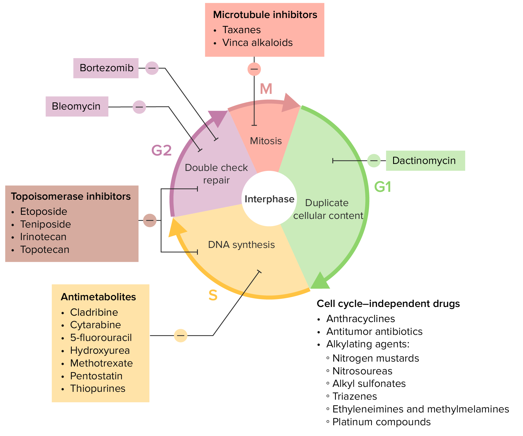
The p53 protein is the product of mutations in the p53 gene. It plays an anti-proliferative effect in different types of stress responses, including cell cycle arrest and apoptosis. For example, when cells are damaged or cell proliferation is abnormal, the p53 gene is activated, leading to cell cycle arrest and even cell apoptosis . In tumor Keywords: cell cycle arrest, DNA damage, chemotherapy. Introduction. The normal cell cycle consists of complex pathways that regulate the duplication of all molecules and organelles and their separation into two identical daughter cells. This progresses through four phases: G1 (gap), S (synthesis), G2 (gap) and M (mitosis) and is coordinated by
Impairment of caspase activity using pan-caspase inhibitor Z-VAD reveals this chemotherapy-induced apoptotic pathway as an essential factor in driving synergy. Mannose-6-phosphate receptor-mediated autophagy and the arrest of cell cycle in G2/M are also shown to be induced by chemotherapy and significantly contributing to the synergy.
