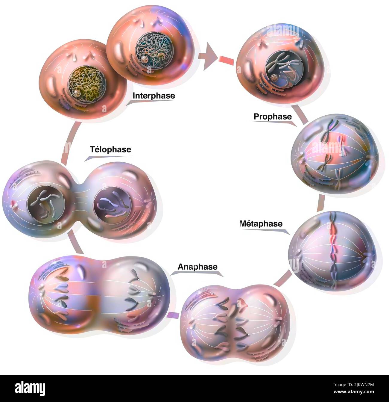
The Stages of Mitosis and Cell Division Biology Diagrams It involves a series of tightly regulated steps, including interphase and prophase. While both interphase and prophase are crucial stages of the cell cycle, they differ in several key attributes. In this article, we will explore and compare the characteristics of interphase and prophase, shedding light on their distinct roles in cell division. What is Prophase. Prophase is the first phase of the mitotic cell division. In meiosis, two prophase stages can be identified: prophase 1 and prophase 2.During the prophase of mitosis, chromatids are condensed into chromosomes, exhibiting short and thick, thread-like structures. Since DNA replication has occurred previously in the interphase, each chromosome contains two identical copies of As living organisms grow, their cells must replicate and divide. Most animal cells, except for sex cells, undergo the process of mitosis to create new cells. Through mitosis, one cell creates two genetically identical daughter cells. Mitosis is a complex process consisting of multiple phases; anaphase, interphase, metaphase and prophase. Each phase has its own steps and is integral to the
:max_bytes(150000):strip_icc()/interphase-58e3d4a45f9b58ef7e071ea0.jpg)
Before a dividing cell enters mitosis, it undergoes a period of growth called interphase. About 90% of a cell's time in the normal cell cycle may be spent in interphase. G1 phase: The period before the synthesis of DNA. In this phase, the cell increases in mass in preparation for cell division. The G1 phase is the first gap phase.
Interphase vs. Prophase - What's the Difference? Biology Diagrams
The material around the nucleoli, contained within the nuclear envelope is DNA in the form of chromatin. This will not pick up a stain well and so will not appear as distinct shapes within the nucleus. Find these indicators of interphase in cell A in the image below. Figure \(\PageIndex{1}\): Cells in an onion root in interphase and prophase.
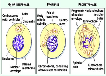
Interphase The cell spends most of its life in this phase. The DNA in chromosomes copies itself ready for mitosis. Prophase The DNA in chromosomes and their copies condenses to become more visible
Edexcel The stages of mitosis in detail Biology Diagrams
Learn the four stages of mitosis, the process of cell division that produces identical daughter cells. Prophase is when chromatin condenses into chromosomes and the spindle apparatus forms. Prophase. Most of the time, DNA is tightly coiled and structured around proteins called histones. This packaged form is known as chromatin. The first stage of mitosis sees this chromatin supercoiling from their operational width of 30nm, to the 500nm thickness associated with chromosomes. Some cells remain in interphase for days or even years; some cells never leave interphase. At the end of interphase, the cell has duplicated its chromosomes and is ready to move them into separate cells, called daughter cells. This occurs during the four steps of mitosis, called prophase, metaphase, anaphase and telophase.
:max_bytes(150000):strip_icc()/interphase-58e3d4a45f9b58ef7e071ea0.jpg)